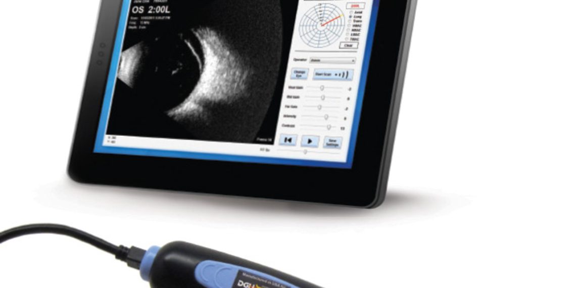
✍️ B-scan echograms are read from left (probe contact with globe) to right (orbital echoes).
✍️ some other scanners, it is read from top to bottom of the echogram.
✍️ In axial scans, the lens and optic nerve are displayed.
✍️ the optic nerve produces a characteristic V-shaped dark shadow in an otherwise bright (high reflective) homogeneous orbital pattern
✍️ In a longitudinal scan, the optic nerve head and anterior portion of the retrobulbar optic nerve are displayed at the bottom of the echogram. At the top is the fundus periphery and ciliary body .
✍️ In L12 and L6 sections, the dark shadow of the superior and inferior recti is displayed in the orbit pattern.
✍️ In L3 and L9, the shadow of the medial and lateral recti is displayed.
✍️ In a transverse scan, the lens and optic nerve shadow are not normally displayed and cross section of a rectus muscle can be seen in its corresponding location
✍️ In normal globe scans no echoes are seen in the vitreous cavity, the normal globe contour is observed, and no membrane or mass lesion is encountered.
✅ Globe lesions that produce an abnormal B-scan can be summarized into four categories:
✍️ Membrane lesion
💧retinaldetachment
💧retinoschisis
💧 choroidal detachment
💧 PVD
💧 persistent hyperplastic primary vitreous.
✍️ Mass lesion
💧melanoma
💧nevus
💧metastasis
💧hemangioma
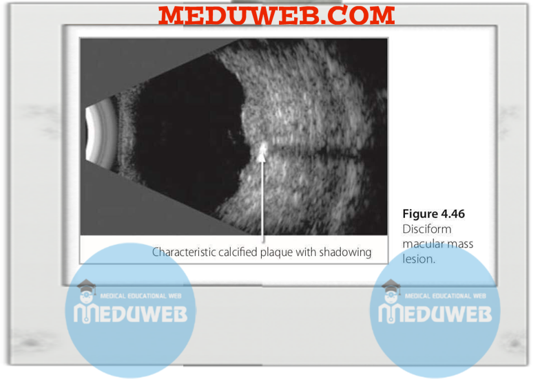
💧macular disciform
💧eccentric disciform lesions
💧tuberous sclerosis
💧inflammatory nodules.
✍️ Discrete opacities
💧asteroid hyalosis
💧VH
💧intraocular foreign body
✍️ Globe wall abnormalities
💧globe deformities
💧staphyloma
💧idiopathic sclerochoroidal calcification
💧choroidal osteoma
💧disc drusen
💧phithisis bulbi.
💧Posterior scleritis
✅ Normal and abnormal B-scan Common Artifacts
✍️inadequate jell
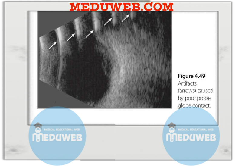
✍️ weak contact between probe and globe
✍️ from IOL implant when scanning axially.
✍️shadowing from calcification
✍️ shadowing from intraocular foreign or retained oil bubbles
✅ Sonographic criteria of common ocular lesions
✍️ Choroidal disorders
👉 Ultrasound is an important investigation in cases of choroidal folds to rule out a retrobulbar orbital mass such as optic nerve swelling
👉an orbital mass such as hemangioma where folds are located at the posterior pole
👉a lacrimal gland tumor where folds are normally discovered at a supratemporal fundus location.
👉 Posterior scleritis is a cause of choroidal folds and is easily diagnosed on ultrasound by characteristic T sign
👉 In many cases, choroidal folds are idiopathic and ultrasound is helpful in ruling out orbital causes.
👉 It will often show flattening of the globe wall and reduction in axial eye length accompanied clinically by progressive hypermetropia over time
✍️ Optic nerve disorders
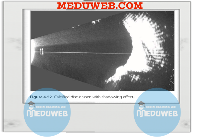
👉a method of choice in the detection of optic nerve head drusen as it shows, very readily, the calcification at the nerve head with its characteristic shadowing effect
👉 It is also a good method for buried nerve drusen.
👉best images of optic nerve head drusen are obtained at a relatively low gain setting to enhance the high reflective, calcified nature of the drusen caused by the high calcium contents.
👉 Care must be taken not to confuse a bright disc papilla with drusen, and the diagnosis of disc drusen must only be made in cases where calcification and shadowing are clearly demonstrated.

✅Intraocular neoplasms
✍️ Choroidal melanoma
👉 a smooth dome or a mushroom-shaped solid mass on B-scan
👉Rarely, it presents as the diffuse melanoma type and appears as a slight irregular thickening of the choroidal layer extending over a wide area of the globe wall
👉 A dome-shaped tumor centered in the beam path may produce the well-known choroidal excavation.
👉 Because of the low reflective nature of melanoma, the characteristic feature of acoustic hollowing has been described.
👉 Less commonly B-scan images can show irregular internal architecture in large tumors and, more rarely, ossification
👉 Melanoma can occupy any part of the fundus and may be associated with retinal detachment, indicating initiation or acceleration of growth.
👉 Careful scanning of the entire tumor base is important to exclude extrascleral extension, a significant prognostic finding that is likely not to be detected on clinical examination andby using other retinal imaging techniques.
👉 Precise localization and measurement of the tumor are undertaken on B-scan by performing transverse, longitudinal, and occasional axial sections.
👉 lateral (base) dimensions are obtained from B-scans, and measurement of the tumor height is performed on well-centered B-scan and more accurately on standardized A-scan.
👉 the precise localization and measurements of the tumor are required by the oncologist to accurately place the scleral plaque and deliver the correct dose of radiation treatment.
👉 A quantitative examination involves A-scan assessment of reflectivity, internal structure, and sound attenuation.
💧Malignant melanoma produces low-to-medium reflective, regularly structured lesion spikes, with medium-to-high sound attenuation.
👉 On kinetic echography, vascularity is common and is diagnostic.
💧On A-scan this appears as fine, fast, vertical oscillation of echoes within the tumor mass.
👉 In very vascular, commonly mushroom-shaped lesions vascularity is also evident on B-scan.
✍️ Choroidal metastasis
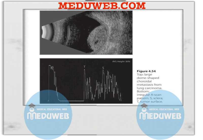
👉 On topographic B-scan evaluation, choroidal metastases appear as dome-shaped lesions but more characteristically as masses with an irregular outline.
👉 they are most frequently located in the posterior pole.
👉 tend to be associated with extensive retinal detachment, a manifestation of rapid growth .
👉 More than one lesion in the same eye and bilateral presentation are not uncommon, and follow-up studies usually showing rapid growth of the tumor over a relatively short period of time (few weeks).
👉 A quantitative examination on A-scan shows a variable reflectivity depending on the primary lesion.
💧 the internal structure is irregular, and the sound attenuation is weak.
👉 Vascularity in metastasis is normally not detected, but it has been rarely reported in some cases of small cell carcinoma of the lung
✍️ Choroidal hemangioma
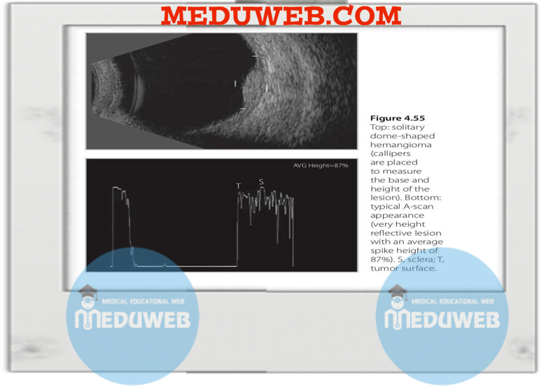
👉 Topographically, choroidal hemangioma appears either as a bright, solitary, dome-shaped, solid lesion located at the posterior pole and around the disc or macula
👉 may be as a diffuse thickening of the choroidal layer that is commonly associated with Sturge–Weber syndrome
👉 Hemangioma tends to remain static or grow very slowly and rarely produces retinal detachment
👉 localized focal cystic retinal changes may be seen in long-standing cases.
👉 Quantitative examination shows a very highly reflective, regularly structured tissue, producing weak sound attenuation.
👉 On kinetic examination, although choroidal hemangiomas are classified as vascular hamartomas, they do not show the fast moving spikes on A-scan, as the large honeycomb spaces are filled with stagnant blood, with no significant blood flow.
🌕Choroidal osteoma
👉 unilateral
👉located at the posterior pole or at the macula.
👉flat (only showing a calcified flat globe wall)
👉slightly elevated in an irregular pattern
👉Scanning of both eyes is recommended in such cases to rule out idiopathic sclerochoroidal calcification that looks similar to choroidal osteoma on ultrasound but presents more commonly as bilateral multiple lesions that are located more peripherally in the globe wall.


